


10 min read
Ankle dorsiflexion is one of the most clinically significant and consistently undertested mobility qualities in fitness and coaching practice. When it is limited, clients cannot perform a safe full-depth squat without compensation.
They develop shin splints, patellar tendinopathy, and Achilles tendinopathy at rates that would be meaningfully reduced with systematic dorsiflexion assessment. They land from jumps with biomechanics that increase their risk of ACL injury. They walk and run with gait mechanics that gradually overload the knee and lower back.
Yet most coaches spend minimal time testing dorsiflexion, fewer still understand what is causing the restriction in a given client, and fewer still know which interventions address the cause rather than just the symptom.
This guide covers the complete picture: precise definition and anatomy, normal ranges and how to measure them, how limited dorsiflexion cascades through the kinetic chain, the causes of restriction, the connection to specific injuries, and the evidence-based exercises and protocols that actually improve it.
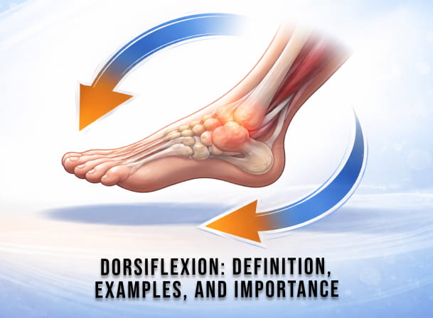
What Is Dorsiflexion?
Dorsiflexion is the movement at a joint that brings the dorsal (top) surface of the foot or hand closer to the limb above it.
At the ankle (talocrural joint), dorsiflexion occurs when the top of the foot moves upward toward the shin, reducing the angle between the dorsum of the foot and the anterior surface of the tibia. This is the motion that occurs when you lift your toes off the floor with your heel planted, when your shin moves forward over your foot during a squat descent, or when your foot clears the ground during the swing phase of running.
At the wrist, dorsiflexion (also called wrist extension) occurs when the back of the hand moves toward the dorsal side of the forearm. This matters in fitness contexts during front rack positioning in Olympic lifting, the bottom of a push-up, and other upper-body loaded positions.
Unless otherwise specified, "dorsiflexion" in the fitness and coaching context refers to ankle dorsiflexion.
Two Types of Ankle Dorsiflexion
Non-weight-bearing (open-chain) dorsiflexion: The foot is lifted toward the shin while the leg is free, as in lying on your back and pulling your toes toward you. This is what goniometric measurement typically captures in clinical settings. Normal open-chain dorsiflexion is approximately 20 degrees past neutral.
Weight-bearing (closed-chain) dorsiflexion: The foot is planted on the ground, and the shin moves forward over the foot, as in a squat, lunge, or stair descent. This is the functionally relevant form for athletic and training contexts. It is assessed with the knee-to-wall (weight-bearing lunge) test and is typically the more limited of the two in clients who have restricted mobility.
Weight-bearing dorsiflexion is more relevant for programming decisions because it limits squat depth, causes compensations in the lunge, and governs landing mechanics.
The Anatomy of Dorsiflexion
Primary Muscle: Tibialis Anterior
The tibialis anterior is the primary producer of ankle dorsiflexion. It runs along the lateral surface of the tibia (shin bone) from its origin at the upper two-thirds of the lateral tibia and interosseous membrane to its insertion at the medial cuneiform and the base of the first metatarsal.
When the tibialis anterior contracts concentrically, it lifts the foot upward (dorsiflexion) and simultaneously inverts the foot (turns the sole inward). When it contracts eccentrically, as during the heel-strike phase of walking and running, it controls the rate at which the foot lowers to the ground, preventing foot slap and absorbing impact.
Weakness of the tibialis anterior is the direct cause of "foot drop," a condition in which the client cannot lift the foot adequately during the swing phase of gait, resulting in toe dragging. Even sub-clinical tibialis anterior weakness contributes to poor shock absorption and reduced active control of dorsiflexion during dynamic movement.
Secondary Muscles: Extensor Hallucis Longus and Extensor Digitorum Longus
The extensor hallucis longus (EHL) primarily extends the big toe but contributes secondary dorsiflexion force through its line of pull anterior to the ankle joint axis. The extensor digitorum longus (EDL) primarily extends toes 2 through 5 and similarly contributes secondary dorsiflexion alongside toe extension.
Both muscles are important for toe clearance during the swing phase of gait. Their weakness contributes to stumbling, toe dragging, and compensatory hip hiking during running.
The Opposing Structures: What Limits Dorsiflexion
Dorsiflexion is passively limited by the posterior calf complex (gastrocnemius and soleus), the Achilles tendon, the posterior joint capsule of the talocrural joint, and the posterior soft tissue structures of the ankle. The specific structure causing restriction has implications for which intervention is appropriate:
Gastrocnemius tightness: Limits dorsiflexion most when the knee is extended (straight), because the gastrocnemius crosses both the knee and ankle. Standing calf stretches with a straight knee address tightness in the gastrocnemius.
Soleus tightness: Limits dorsiflexion regardless of knee position because the soleus only crosses the ankle. Bent-knee calf stretches and seated stretches specifically address soleus tightness.
Posterior joint capsule restriction: Limits dorsiflexion mechanically rather than through muscle stiffness. Responds to joint mobilization and banded distraction techniques rather than stretching.
Bony impingement: An osteophyte or bony spur at the anterior ankle joint can create a hard end-feel during dorsiflexion. This requires medical evaluation and does not respond to stretching.
Normal Dorsiflexion Range of Motion
Understanding normal ranges is essential for evaluating clients and identifying meaningful restrictions.
Non-weight-bearing ankle dorsiflexion: Approximately 20 degrees of dorsiflexion past neutral (the position with the foot at a right angle to the tibia), with total passive dorsiflexion reaching 20 to 30 degrees in most healthy adults.
Weight-bearing dorsiflexion during gait: At least 10 degrees of weight-bearing dorsiflexion is required for efficient walking mechanics. More recent gait analysis research has reported that up to 20 degrees of dorsiflexion may occur during normal walking.
Weight-bearing dorsiflexion during deep squatting: A 2006 study published in the Journal of Orthopaedic Research found that squatting to just past parallel requires approximately 35 degrees of ankle dorsiflexion. The deeper the squat, the greater the demand: a full below-parallel squat places the ankle at the limit of its dorsiflexion range in most people.
Knee-to-wall test reference: In the weight-bearing lunge test (half-kneeling dorsiflexion test), touching the knee to a wall with the big toe 5 inches from the wall represents adequate ankle dorsiflexion for most athletic activities. Being unable to touch the wall at 3 to 4 inches indicates a clinically relevant restriction.
How to Assess Dorsiflexion
Coaches do not need clinical measurement tools to screen for meaningful dorsiflexion restriction. Two practical tests cover most coaching needs.
Knee-to-Wall Test (Weight-Bearing Lunge Test)
This is the most reliable, standardized field assessment for weight-bearing dorsiflexion. It is used across physical therapy and strength and conditioning research and can be performed without equipment.
Setup: Stand or kneel barefoot facing a wall. Place the big toe of the test foot at a measured distance from the wall (start at 4 to 5 inches). Keeping the heel flat on the ground, drive the knee forward toward the wall, directly over the second toe.
Pass criteria: The knee touches the wall at 5 inches without the heel lifting, the foot rolling inward (pronating), or the knee deviating medially or laterally.
Fail criteria: The knee cannot reach the wall at 4 inches, or it reaches the wall only with compensatory heel rise, foot rotation, or knee valgus.
Bilateral comparison: Always test both sides. A difference of 1-2 cm or more between sides indicates asymmetric restriction and warrants targeted intervention on the more restricted side.
Overhead Squat Assessment
The overhead squat assessment, which is part of a complete personal training assessment protocol, reveals dorsiflexion restriction indirectly through the compensatory patterns it produces:
Heel rise: Heels lift off the ground during the descent, indicating insufficient dorsiflexion range for the squat depth being attempted.
Forward trunk lean: Excessive forward inclination of the torso is a common compensatory mechanism for dorsiflexion. The hips shift backward to maintain balance when the ankle cannot allow the shin to travel forward, and the trunk follows.
Foot flare (external rotation): Feet rotating outward is one of the most common dorsiflexion compensations. External rotation increases the effective dorsiflexion range available by changing the plane in which the shin moves forward.
Knee valgus (inward collapse): Limited dorsiflexion increases ground reaction forces and has been consistently linked to dynamic knee valgus during squatting, landing, and step-down tasks, which are primary biomechanical risk factors for ACL and patellar tendon injuries.
Why Limited Dorsiflexion Matters: The Kinetic Chain Cascade
The ankle is the foundation of the lower extremity kinetic chain. An ankle restriction forces compensatory motion at every joint above it, creating a cascade of movement dysfunction that travels upward through the knee, hip, and lower back.
Squat and Lunge Mechanics
The most immediately visible consequence of limited dorsiflexion in a training context is compromised squat mechanics. When the ankle cannot produce sufficient dorsiflexion range, one or more of the following compensations always occur: heel rise, forward trunk lean, foot flare, or knee valgus. Each compensation transfers load to structures not designed to bear it in that pattern.
A client who cannot achieve adequate dorsiflexion for deep squatting does not need to be coached harder on squat form. They are a client who needs ankle mobility work before the squatting depth is progressed. Loading a compensated squat pattern increases injury risk rather than building strength.
Running and Gait
During the gait cycle, adequate dorsiflexion is required in two distinct phases. In the swing phase, the foot must dorsiflex to clear the ground. In the stance phase, particularly at mid-stance and late stance, the tibia must travel forward over the planted foot, requiring controlled, progressive dorsiflexion.
Limited dorsiflexion during stance results in an early heel rise (the heel lifts prematurely before toe-off), altering the timing and mechanics of push-off. Compensations in gait propagate upward: clients with restricted dorsiflexion often display excessive knee flexion at initial contact, altered hip extension patterns, and lumbar extension compensation.
Landing Mechanics
Research consistently links limited ankle dorsiflexion to increased ground reaction forces during landing. When the ankle cannot absorb impact through adequate dorsiflexion, the knee and hip must absorb proportionally more of the force. The same research links reduced ankle dorsiflexion to increased knee valgus during landing, which is the primary biomechanical antecedent to non-contact ACL tears.
A 2011 study in the Journal of Athletic Training found direct associations between restricted ankle dorsiflexion range and adverse landing biomechanics, including reduced knee flexion at initial contact and increased knee valgus angles. These findings have direct implications for coaches working with athletes in any sport that involves jumping and landing.
Dorsiflexion and Injury Risk
The research on dorsiflexion restriction as an injury risk factor is consistent and clinically significant. Coaches who do not assess it are missing one of the most modifiable risk factors in their client populations.
Achilles Tendinopathy
Reduced ankle dorsiflexion forces the gastrocnemius and soleus to absorb greater proportional load during activities requiring plantar flexion. When dorsiflexion is limited, force absorption that would normally be distributed across the ankle joint complex is transferred to the Achilles tendon. Research cited in the Electronic Journal of General Medicine found that decreased ankle dorsiflexion was associated with a 2.5- to 3.6-fold greater risk of Achilles tendinopathy. This relationship is particularly relevant in runners and athletes with high volumes of repetitive plantar flexion loading.
The FitBudd resource on plantar flexor muscles covers the calf complex anatomy and training implications that interact directly with dorsiflexion restriction in injury prevention programming.
Patellar Tendinopathy
A study published in the Journal of Science and Medicine in Sport found that reduced ankle dorsiflexion range increased the risk of patellar tendon injury among volleyball players. The mechanism is consistent with the kinetic chain model: when dorsiflexion is restricted, the knee must compensate during landing and deceleration, increasing patellofemoral joint stress. The compensatory knee valgus associated with restricted dorsiflexion also increases medial patellar contact pressure.
ACL Injury
Research has established that restricted ankle dorsiflexion is associated with dynamic knee valgus during functional movements including squatting, single-leg squatting, lateral step-down, and landing from jumps. Since dynamic knee valgus is a primary mechanism for non-contact ACL tears, the link between restricted dorsiflexion and ACL injury risk is a significant concern for coaches working with athletes.
Plantar Fasciitis
Tight calf muscles, secondary to limited ankle dorsiflexion, increase tension on the plantar fascia via the windlass mechanism. As the calf complex becomes shorter and stiffer, it places more traction on the plantar fascial insertion at the calcaneus. Calf stretching, which directly increases dorsiflexion range, is consistently among the most effective first-line interventions for plantar fasciitis.
Shin Splints (Medial Tibial Stress Syndrome)
The tibialis anterior, the primary dorsiflexor, is also the muscle most commonly implicated in shin splints due to eccentric overload during the foot-strike phase of running. Weakness in the tibialis anterior and restricted dorsiflexion range both contribute to altered gait mechanics that increase tibial stress. Strengthening the tibialis anterior, along with improving calf flexibility, addresses both the muscle and mobility contributors simultaneously.
Causes of Restricted Dorsiflexion
Understanding why a client's dorsiflexion is limited helps determine which intervention will address it.
Chronic calf muscle tightness: The most common cause. Results from sedentary behavior, prolonged sitting with the ankle in slight plantar flexion, consistent use of high-heeled footwear, and high-volume calf training without adequate stretching. Responds well to targeted calf stretching and foam rolling.
Previous ankle sprain: Lateral ankle sprains that have not been fully rehabilitated commonly leave the posterior joint capsule contracted and fibrotic. This produces a joint-based restriction that does not respond to muscle stretching. Responds to joint mobilization, distraction techniques, and weight-bearing mobility work.
Scar tissue and joint capsule restriction: Following any ankle injury, surgery, or immobilization, intra-articular adhesions and capsular tightening can mechanically restrict dorsiflexion. The end-feel is firm and often asymmetric compared to the unaffected side.
Footwear habits: Extended daily use of elevated-heel shoes (including athletic shoes with significant heel drop) habitually shortens the calf complex. Transitioning to lower-heeled footwear and gradually increasing barefoot exposure can contribute to long-term improvement in dorsiflexion.
Genetics and bony anatomy: Some individuals have naturally less talocrural joint mobility due to the shape of the talus and tibial plafond. This is a genuine structural limitation that cannot be changed through stretching, though training in greater ranges can improve functional control of the existing range.
Exercises to Improve Dorsiflexion
1. Kneeling Ankle Dorsiflexion Stretch (Knee-to-Wall Mobilization)
Purpose: The most effective weight-bearing dorsiflexion mobility exercise. Stretches the posterior calf complex and trains the ankle through its weight-bearing range simultaneously.
Execution: Kneel in a half-kneeling position facing a wall. Place the front foot flat on the floor approximately 3 to 4 inches from the wall. Drive the knee forward directly over the second toe, attempting to touch the wall while keeping the heel planted. At the limit of range, add gentle overpressure with the hand on the knee. Hold 2 seconds at end range, return, repeat.
Programming: 3 sets of 10 to 15 reps per side, performed before training sessions involving squats, lunges, or running. Can be used daily without recovery concerns.
Progression: Gradually increase the distance from the wall as range improves. Track improvement objectively using the knee-to-wall test measurement.
2. Standing Gastrocnemius Stretch
Purpose: Targets the gastrocnemius specifically. Most effective for clients whose restriction is worse with a straight knee, indicating gastrocnemius tightness as the primary limiting factor.
Execution: Stand facing a wall with your hands for support. Step one foot back approximately 60 to 90 cm. Keep the back knee fully extended. Press the back heel into the floor and lean forward until a stretch is felt in the upper calf. Hold 30 to 60 seconds. Switch sides.
Programming: 3 holds of 30 to 60 seconds per side. Best performed post-workout as part of a cool-down, or during dedicated mobility sessions. Not recommended immediately before heavy strength training due to the potential for a transient reduction in force production.
3. Seated Soleus Stretch (Bent-Knee Calf Stretch)
Purpose: Targets the soleus specifically. Essential when restriction is present regardless of knee position, indicating the soleus rather than the gastrocnemius as the primary limiting structure. This is a commonly undertrained stretch because the soleus is easily overlooked in standard flexibility work.
Execution: Sit on the floor or on a low surface with one knee bent at approximately 90 degrees. Keep the foot flat on the floor. Lean forward, pressing the knee gently toward the toes. Hold 30 seconds. Alternatively, stand with a slight bend in the knee and press forward into a wall.
Programming: 3 holds of 30 to 60 seconds per side. The seated position makes this accessible for almost all populations, including older adults and clients with balance limitations.
4. Banded Ankle Distraction
Purpose: Addresses posterior joint capsule restriction and bony tightness that does not respond to muscle stretching. The distraction force shifts the talus posteriorly, creating space for dorsiflexion that tightness in the joint capsule normally prevents.
Execution: Anchor a resistance band to a fixed low point. Place the loop around the front of the ankle just above the talus. Step back to create tension that pulls the ankle forward (anteriorly). From a half-kneeling position with the banded ankle in front, drive the knee forward into dorsiflexion while the band provides posterior distraction. Hold 2 to 3 seconds at the end range, return.
Programming: 3 sets of 10 to 15 reps per side. Particularly useful for clients with a history of ankle sprains or those who show little improvement with static stretching alone. Should be used before training sessions.
5. Tibialis Anterior Strengthening (Resistance Band Dorsiflexion)
Purpose: Directly strengthens the primary dorsiflexion muscle. Prevents shin splints, improves foot clearance during gait, and builds eccentric control during heel-strike. This is often overlooked in favor of passive mobility work when in fact active muscle strength is what determines functional dorsiflexion control.
Execution: Sit on a bench or chair with the legs extended. Anchor a resistance band to a fixed point in front of you and loop it around the top of the foot. Dorsiflex the foot (pull toes toward shin) against the band resistance. Control the return slowly. 3 sets of 15 to 20 reps.
Variation: Walking on heels (tibialis anterior activation through gait) for 30 seconds per set is an effective low-tech alternative.
Programming: 2 to 3 times per week, particularly in clients presenting with shin splints, foot drop, or poor active dorsiflexion control despite adequate passive range.
6. Heel Drops (Eccentric Calf Raises)
Purpose: A dual-function exercise that simultaneously strengthens the calf complex and improves dorsiflexion range through the eccentric loaded stretch at the bottom of the movement. Particularly effective for Achilles tendinopathy rehabilitation and prevention.
Execution: Stand on the edge of a step with the balls of both feet on the step edge. Rise onto both toes using both legs. Transfer to one leg. Slowly lower the heel below the step edge over 3 to 5 seconds. Step down and repeat.
Programming: 3 sets of 15 reps per side. The slow eccentric phase is the critical component. Progress by adding a loaded dumbbell as strength improves.
7. Toes-Elevated Squats
Purpose: A dynamic warm-up drill that allows clients with restricted dorsiflexion to train squat mechanics through a fuller range by reducing the dorsiflexion demand. Simultaneously provides active dorsiflexion training within the squat pattern.
Execution: Place the toes and ball of the foot on a 1 to 1.5-inch elevation (a small weight plate works well). Keep heels on the floor. Perform bodyweight squats through the fullest comfortable range. The elevation reduces the dorsiflexion requirement, allowing deeper squat depth while the mobility limitation is being addressed.
Programming: Use as a warm-up modification while actively working on ankle mobility. Do not use it permanently as a substitute for developing actual dorsiflexion range.
Dorsiflexion Programming for Coaches
Assess first: Do not prescribe dorsiflexion work without understanding the cause of restriction. Use the knee-to-wall test on every new client. Compare bilaterally. Note whether the restriction improves with the knee bent (suggesting gastrocnemius tightness) or not (suggesting soleus or joint-based restriction). This determines which intervention is most appropriate.
Address the cause, not just the symptom: Clients with joint-based restriction from previous ankle sprains need banded distraction and joint mobilization, not more calf stretching. Clients with gastrocnemius tightness need straight-knee stretching. Clients with poor tibialis anterior strength need active dorsiflexion strengthening, not just passive mobility work.
Integrate mobility into sessions, not separately: Dorsiflexion mobility work done immediately before squatting or lunging provides both mobility benefit and warm-up effect. A knee-to-wall mobilization protocol before each squat session produces cumulative improvement without requiring separate mobility sessions.
Track improvement objectively: Reassess the knee-to-wall test distance every 4 to 6 weeks. Document measurements for each client. Improvement of 1 to 2 cm per month with consistent mobility work is realistic. If no improvement after 8 weeks of targeted intervention, consider whether the restriction is joint- or soft-tissue-based.
Modify training immediately: Do not wait for dorsiflexion to improve before modifying training. Use toes-elevated squats, reduce squat depth to a range the client can control without compensation, or substitute exercises that require less dorsiflexion (e.g., leg press, split squats with a lower ROM) while the mobility is being developed.
Dorsiflexion assessment belongs in every personal trainer assessment protocol alongside overhead squat assessment, hip mobility screening, and thoracic rotation testing. The FitBudd guide to corrective exercise provides the broader corrective programming framework within which dorsiflexion work fits alongside other movement quality interventions.
For the counterpart of this article covering the opposing ankle motion and how plantar flexion and dorsiflexion work together across the complete range of ankle function, see the FitBudd guide to dorsiflexion vs plantar flexion.
Conclusion
Dorsiflexion is not a peripheral mobility concern. It is a foundational movement quality that determines the safety and efficiency of every major lower body pattern a coach programs: squats, lunges, running, jumping, and landing. Limited dorsiflexion leads to compensations at every level of the kinetic chain above the ankle, and it is a modifiable risk factor for some of the most common and debilitating lower-extremity injuries in training populations.
The coaching implication is clear: assess dorsiflexion on every new client using the knee-to-wall test, identify whether restriction is soft-tissue or joint-based, prescribe the correct intervention, modify training immediately to accommodate the restriction, and track improvement over time.
FitBudd makes it straightforward to document ankle mobility assessments, build corrective dorsiflexion protocols directly into client training plans, and track improvement across reassessments over time. Start your free 30-day trial at FitBudd and build the assessment infrastructure that separates coaching based on evidence from coaching based on guesswork.
%20to%20Become%20a%20Certified%20Personal%20Trainer-min.jpg)


